Photobucket
Photobucket

HELLO <body><script type="text/javascript"> function setAttributeOnload(object, attribute, val) { if(window.addEventListener) { window.addEventListener('load', function(){ object[attribute] = val; }, false); } else { window.attachEvent('onload', function(){ object[attribute] = val; }); } } </script> <div id="navbar-iframe-container"></div> <script type="text/javascript" src="https://apis.google.com/js/platform.js"></script> <script type="text/javascript"> gapi.load("gapi.iframes:gapi.iframes.style.bubble", function() { if (gapi.iframes && gapi.iframes.getContext) { gapi.iframes.getContext().openChild({ url: 'https://www.blogger.com/navbar.g?targetBlogID\x3d4645296235723893839\x26blogName\x3dMicrobiology+B\x26publishMode\x3dPUBLISH_MODE_BLOGSPOT\x26navbarType\x3dBLUE\x26layoutType\x3dCLASSIC\x26searchRoot\x3dhttps://microvirology.blogspot.com/search\x26blogLocale\x3den\x26v\x3d2\x26homepageUrl\x3dhttps://microvirology.blogspot.com/\x26vt\x3d6711310578554945636', where: document.getElementById("navbar-iframe-container"), id: "navbar-iframe" }); } }); </script>
Tuesday, January 27, 2009

Picornaviridae

It is the largest virus family over 230 serotypes.
It can be classified into 5 genera :


Enterovirus
Human polioviruses 1-3 (serotypes=3)
Human hepatitis virus A (serotypes=1)

Rhinovirus
Human rhinoviruses 1-100 (serotypes=100)
Bovine rhinoviruses 1-2 (serotypes=2)

Aphthovirus
Foot-and-mouth disease virus 1-7 (serotypes=7)

Cardiovirus
Encephalomyocarditis (EMC), Mengo (serotypes=1)

Unassigned
Equine rhinoviruses 1-2, Cricket paralysis... (serotypes>3)


Morphology

Virions consist of a capsid. Virus capsid is not enveloped, round with icosahedral symmetry. The capsid is isometricand has a diameter of (22-)27-30 nm. Capsids appear round. The capsid consists of 12 capsomers.
Incomplete particles are common. They are devoid of nucleic acid characterized by capsids with dark centers in negative stain preparations.


Genome of Picornaviridae

Poliovirus virion RNA functions as an mRNA but does not have the methylated cap structure typical of eucaryotic mRNAs - it has a internal ribosome entry site or IRES which enables ribosomes to bind without having to recognize a 5' methylated cap structure.


The mRNA is translated into a single polypeptide (polyprotein), which is cleaved. The cleavages occur before translation is complete and are carried out by virally coded proteases.


Example of Picornaviridae: Rhinovirus (Common Cold)

Pathogenesis
- Infect the lower respiratory tract
- Trigger asthma exacerbation
- Short incubation of (2 -3 days)
- Over 100 Rhinoviruses.
- Mild upper respiratory viral infection


Symptoms
- Watery nasal discharge
- Congestion
- Sneezing
- Little / No fever


Preventions


Example of Orthomyxoviridae: Influenza (Flu)


Diseases related to respiratory system are spread more in crowded environment and enclosed spaces, such as buses. Transmission may also occur by direct contact, since the influenza virus may survive for hours, particularly in cold and in low humidity.

Morhology
Virions enveloped , pleomorphic and filamentous forms occur; spherical, or filamentous; 50-120 nm in diameter, or 20 nm in diameter; 200-300(-3000) nm long. Surface projections of envelope distinct; about 500 spikes (projecting 10-14 nm from the surface); dispersed evenly all over the surface.


Pathogenesis
The influenza A virus can infect humans, horses, birds, and pigs. Wild birds are the natural reservoir for influenza A viruses. The Influenza A virus is mainly spread by aerosols that get into the respiratory tract. The virus particles then attack the ciliated epithelial cells lining the respiratory tract, causing necrosis of the cells.


Prevention


Laboratory Diagnosis


Differences between Cold and Flu


Video of how influenza virus affect a human body!


Video of mutation of an antigenic shift in human genetic code!


**ALL IMAGES CLICK TO ENLARGE !!

Applications
http://pathmicro.med.sc.edu/mhunt/RNA-HO.htm
http://www.ncbi.nlm.nih.gov/ICTVdb/ICTVdB/00.052.htm
http://www.flufacts.com/about/cold.aspx
http://www.flufacts.com/treatment/default.aspx
http://www.virology.net/Big_Virology/BVRNAortho.html
http://www.youtube.com/watch?v=fW_YoQYqUzk
http://www.youtube.com/watch?v=01qwLckBLSM&feature=related


HELLO

5:09 PM

Sunday, January 25, 2009

Herpesviridae

There are many types of viruses grouped under herpesviridae and can be categorised into Alpha, Beta and Gamma herpes. In this blog entry Herpes Simplex 1 and Herpes Simplex 2 viruses under the alpha herpes category, the cytomegalovirus under the Beta herpes category and the Epstein Barr Virus under the gamma herpes category will be discussed.
The diagram below shows the different categories of viruses. However, not all herpes viruses are shown below.

(click image to enlarge)


Features of Herpesviridae Viruses

The unique feature of the herpesviridae viruses’ properties is its ability to stay latent after primary infection and reactivate due to certain factors (stress, menstruation, long exposure to sunlight). After primary infection it stays latent in the nerves cells of the host.
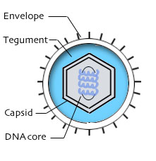
One unique structural feature is its concentric virion which consists of:


· Inner core
· Icosahedral capsid
· Amphorous tegument
· Glycoprotein envelope
· Linear double stranded DNA


Herpes Simplex 1 Virus

The herpes simplex 1 virus causes cold sores and blisters which can affect the mouth, eyes and gums. After primary infection, the virus stays latent in the nerves cells of the host where it can avoid destruction by immune cells until certain factors (stress, menstruation, long exposure to sunlight) reactivates the virus. This reactivated virus causes symptoms such as cold sores.


Almost all of the adult population has contracted the Herpes Simplex 1 virus through kissing and close proximity to one another. Hence to prevent contracting this, avoid kissing and close proximity to people you do not know. However this virus is not life threatening so it is not much of a threat to public health.


Herpes Simplex 2 Virus

The herpes simplex 2 virus or genital herpes virus causes blisters and burning sensations at the genitals. A foul smelling discharge will be produced from 1 to 3 weeks of the infection.

In the United States about 20% of the adult population has contracted the herpes simplex 2 virus. Since this virus is spread through sexual contact, the most effective method to prevent contracting it is to stop any risky sexual behaviour.

Varicella Zoster Virus

The Varicella Zoster Virus causes 2 types of infections when it infects a host. During the initial infection, it causes chicken pox. After the chicken pox has been cured, it stays latent in the nerve tissues to avoid destruction by immune cells until the efficiency of the immune system dimishes (mostly due to old age) causing reactivation. In this reactivation the symptoms manifests itself in the form of shingles.

The symptoms for the primary infection (chicken pox) are shown below:

· Fever
· Lesion on the body
· Scaring
· Can cause damage to the foetus in pregnant women
· Can be passed from mother to child in pregnant women.
· May cause Guillain Barre syndrome


In the secondary infection which occurs due to virus reactivation shingles on the body will form.

Varicella zoster virus infections can be prevented by administering a vaccine which is available and avoiding infected people.

Epstein Barr Virus

This virus affects the nasopharynx pathways and salivary gland and causes the following symptoms:

· Sore throat lasting a few weeks
· Swollen lymph nodes
· Persistent fever
· Fatigue

The Epstein Barr Virus also plays a part in the emergence of 2 types of cancer which are Burkitt’s lymphoma and nasopharyngeal carcinoma.


Cytomegalovirus

The cytomegalovirus is a virus that causes asymptomatic infection in otherwise healthy individuals. In immunocompromised hosts, the viruses causes eye, brain, intestinal and respiratory infections. In newborns it can cause asymptomatic infection to acute symptoms such as fever, brain damage and even death. In adults it can also cause acute symptoms similar to the Epstein Barr Virus.

To prevent contracting the cytomegalovirus, avoid infected persons and avoid blood and bodily fluid contact.


Lab Diagnosis

Diagnosis can be done use blood test, virus cultures and antigen testing to detect the herpes viruses.


Videos

This video shows what happens when varicella zoster virus re-activates after a person has suffered from chicken pox.


References

Pictures:

  1. http://stdgen.northwestern.edu/stdgen/bacteria/hhv2/herpes.diagram.jpg
  2. http://z.about.com/d/dermatology/1/0/J/5/HerpLip.jpg
  3. http://www.hfreedomnetwork.org/pb/wp_a6802eba/images/img1375543abab7c86d3e.jpg
  4. http://www.childhoodhealth.com/images/varicella_large.jpg
  5. http://medicalimages.allrefer.com/large/chicken-pox.jpg
  6. http://microvet.arizona.edu/Courses/MIC419/ToolBox/elisa3.jpg
  7. http://upload.wikimedia.org/wikipedia/commons/7/76/Plaque_assay_macro.jpg

Information:

  1. http://www.healthscout.com/ency/68/44/main.html
  2. http://www.healthscout.com/ency/68/552/main.html


HELLO

5:01 PM


Hepadnaviridae

Hepadnaviridae is a family of viruses that causes liver infections in humans and animals hence the name 'hepadnaviridae' where 'hepa' is the Latin word for liver. Some animals that can be infected with this family of viruses are ducks, birds, wooly monkeys, squirrels and many more.


Features of Hepadnaviridae viruses

The genome of the hepadnaviridae viruses contains a partial double stranded DNA with overlapping reading frames. As a result it is considered to be an RNA intermediate. Due to these properties, hepadnaviridae viruses are grouped under group VII (RNA reverse transcribing viruses) of the Baltimore classification scheme.


(Click to thumnail to view image)
Hepadnaviridae viruses also possess a spherical viral envelope made up of cellular lipids, glycoproteins and surface antigens and an icosahedral capsid.


Hepatitis B: Pathogenesis


The specific hepadnaviridae virus being discussed in this section will be the Hepatitis B virus (HBV). This virus is important due to its ability to infect humans and it being able to cause potentially life threatening liver infection.


About an estimated 600 000 people die each year due to it and 25% of adults who are chronically infected during childhood will die from liver cirrhosis. Upon infection, 90% of neonates, 50% of children and 5-10% of adults will develop chronic hepatitis B. The diagram below shows areas in the world that are at risk of infection.

(click image to enlarge)

The hepatitis B virus targets the liver cells and can cause the following symptoms during an acute infection:

  • yellowing of the skin and eyes (jaundice),
  • dark urine,
  • extreme fatigue,
  • nausea,
  • vomiting and
  • Abdominal pain.

(click image to enlarge)

Following an acute infection, chronic infection can occur which can result in liver cirrhosis and liver cancer. People with chronic hepatitis are still infectious and may not display any symptoms.

There are many types of hepatitis infections which can be grouped into chronic and acute infections. Chronic active hepatitis is the one that causes liver cirrhosis and liver cancer.The other types of hepatitis infections are shown below.


Hepatitis B: Transmission


The hepatitis B virus can be spread from person to person by various means such as:

  • blood contact
  • bodily fluid contact
  • from mother to baby at birth
  • early childhood infections
  • unsafe injection practices
  • sexual contact

The hepatitis B virus is very infectious and can survive outside the body for 7 days and has an average incubation period of 90 days.

Hepatitis B: Prevention

The hepatitis B infections can be prevented by avoiding risky sexual behavior and sharing of needles since it can spread via blood and bodily fluid contact. Moreover, there is a hepatitis B vaccine available which can prevent hepatitis B infections.


Hepatitis infections have been reduced over the years as since 1987 all babies are vaccinated against it.

Hepatitis B: Lab Diagnosis

Patients can be tested for the Hepatitis B virus using serological methods or liver biopsy.

Serological Methods

Under the serological methods, the blood serum can be tested for detectable hepatitis B virus surface antigen, envelope antigen in acute infections and IgM antibodies.

IgM antibodies are antibodies produced during the acute infection and IgG are the antibodies that persist after the acute infection to serve as the 'memory' which will help fight off subsequent acute infections. After an acute infection, IgM antibodies will start to diminish and the titer of IgG antibodies will start to increase.

The presence of these antibodies and antigens can be detected using an ELISA or blood test.

Liver Biopsy

The other method of diagnosing hepatitis B infection is using liver biopsy when tests using serological methods are positive. This is done to confirm the HBV infection and to assess the patient's condition and decide the treatment for the infection. Liver biopsy is done by removing a small piece of the person's liver which will be examined and tested.

(click image to enlarge)

Types of Hepatitis Viruses

The table below shows the different types hepatitis viruses and the modes of transmissions.


Videos

A Hepatitis information video which outlines the symptoms, transmission, prevention, diagnosis and treatment of hepatitis B.


References

Pictures:


  1. http://www.stanford.edu/group/virus/hepadna/2004tansilvis/hep%20b%20genome.gif
  2. http://www.who.int/entity/ith/maps/hepatitisB2007.jpg
  3. http://ontariohepatitistreatment.com/devel2/images/How_a_Biopsy_is_Performed.jpg
  4. http://www.phoenixpeptide.com/catalog/upload/pnxhighlights/pnxhighlights_000000056/notes/ELISA2.gif
  5. http://sg.wrs.yahoo.com/_ylt=A0S0zu7UFnxJ7msBPQMu4gt./SIG=135jkvjtj/EXP=1232955476/**http%3A//www.bioserv-diagnostics.com/images/jpg%2520rgb/AOA_ELISA_IG_typing_rgb.jpg
  6. http://www.mydietsolutions.com/mds/components/com_virtuemart/shop_image/product/8855aecf720f86ebce2c38801634727f.jpg
  7. http://medicalimages.allrefer.com/large/jaundice.jpg

Information

  1. http://www.stanford.edu/group/virus/hepadna/2004tansilvis/Basics.htm
  2. http://www.who.int/mediacentre/factsheets/fs204/en/


HELLO

3:58 PM