

References:
Virus-Host Interactions
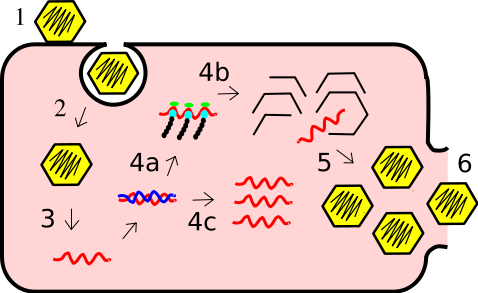
Uncoating
The uncoating of a nucleocapsid which release the viral RNA
Uncoating is an intracellular step during which viral nucleic acid and capsid are separated.
The virus genome is therefore released into the cell.
The virus genome therefore depending, might undergo reverse transcription where the RNA would be converted to DNA.
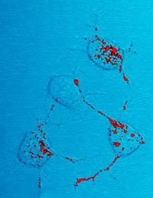
The DNA would then be integrated by the process of integration into the cell’s nuclear
Image above show a HIV’s DNA integrating into a nuclear of a white blood cell’s nuclear.
The DNA is mostly integrated into the cell together with a integrase.
The integrase would assist the viral DNA to be inserted into the cell’s own DNA.
Viral Replication
The replication of HIV virus mRNA.
Since part of the cell’s DNA contains the virus DNA, the cell would also produce virus mRNA together with its normal mRNA.
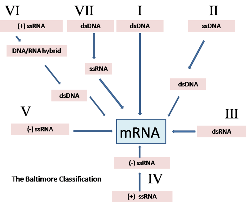
Different Class of virus replicate differently.
There are seven different class of classification.
Class I
This involves double-stranded DNA.
The replication is exclusively nuclear therefore its very dependent on host cell factors
The replication takes place in the cytoplasm, the viral genome contains all factors for genome replication and transcription.
Examples include Adenovirus, Herpesviruses and Poxiviridae.
These virus may cause cancer as they force the cells to undergo cell division.
Class II
This involves single-stranded DNA.
The replication of virus genome takes place in the nucleus
New single-stranded is made from double-stranded DNA formed.
Examples include Parvoviridae and Circoviridae
Class III
This involves double-stranded RNA
The replication of virus takes place all in the cytoplasm
The genome of the virus is fragmented, which mean each different genome code for a different protein.
The replication is monocistronic.
Class IV
This involves single-stranded positive RNA
There are 2 groups, group 1 and group2
Group1
The virus is with polycistronic mRNA, the genome RNA is responsible for forming the mRNA.
These mRNA are later translated into polyprotein.
Therefore from the same strand of RNA, the virus has different method to produce protein.
Group2
This involve virus with complex transcription process.
There is 2 round of transcription before the formation of genomic RNA.
Ribosome frame shifting and proteolytic could be used to produce protein from the same strand of RNA.
Class V
Contains 2 group.
Group1
Virus with non-segmented genome.
The transcription of the negative RNA is induced by RNA-dependent RNA polymerase which gives monocistronic mRNA.
Replication takes place within the cytoplasm.
Group2
Virus with segmented genome.
Replication takes place in nucleus.
RNA-dependent RNA polymerase produce monocistronic mRNA from each segmented genome.
The biggest different between both group is the location where replication takes place.
Class VI
This involve single-stranded positive RNA, but there is DNA intermediate.
It is diploid
The process of reverse transcription of viral RNA to dsDNA is done by viral RT
The dsDNA is integrated into the host cell’s genome
The viral RNA is not used as mRNA
Well studied examples are HIV
Class VII
This involves double stranded DNA, but there is RNA intermediate
Involves overlapping reading of frames
Examples include Hepadnaviriade
This class is not well studied
Assembly or maturation
The assembly of HIV in a white blood cell
Once the various viral subunits have been produced and processed, they must be separated for the final assembly into new virus. This separation, or cleavage, is accomplished by the viral enzyme.
The viral RNA is now repacked into a nucleocapsid again.
The structural subunits of the virus would mesh with a part of the cell membrane causing it to deform.
Release
This is the final step in virus replication, the genetic material enclosed in the nucleocapsid merges with the deformed cell membrane to form a new virus envelope. The virus is therefore released and readily attaches to another cell.
Regulation of expression
Transcriptional Control
· Involve the promotion of viral genome
· Involves the early and later activators / enhancers
· Involves late repressors
· The viral transciptases for RNA viruses is not very understood
Post-transcription control
· Involves the splicing of polycistronic mRNA in nucleus
· There is different rate of splicing
· Involves the control of mRNA from nucleus to cytoplasm
· There are regulatory sequences found on introns
Translational control
· There is different stability of mRNAs
· The secondary structures are close to initiation sequence
· There is problem due to overlapping reading of frames
Translational control – overlapping reading frames
· Internal ribosomal entry sites ( IRES )
· Frameshifting
· Pseudoknots
How could a virus enter a host?
· The skin when there are cuts, abrasions, open wounds
· The eyes (eyelid)
· The lungs when breathed in
· The small intestine, when virus is consumed
· The genitals, during sexual intercourse
Types of virus spread inside a host
· Systemic infection – infect several organs
· Haematogenous spread – spread through the bloodsteam
· Neural spread
Virus could be transmitted
· eating infected tissue (beef, mad cow disease)
· coughing / sneezing ( mucus / saliva )
· contaminated hands ( holding hands / unknowingly touching infected people or items and rub eyes or nose
· Saliva ( kissing , sharing of food )
· Blood ( sharing of needles )
· Sexual intercourse
· Faeces( only affect undeveloped countries as people relieve themselves in river and obtain drinking from river)
Virus induced injuries (CPE)
· Cell death
· Mutation ( 2 nucleus , changed in shape )
· Change in cell membrane permeability
(Others)
· Shut down of the cellular function of the cell which leads to cell death ( example neurons, shut down lead to a person being paralyzed)
· Immunopathological lesions ( HIV, the white blood cell are unable to function. Enhancement in immune response causing haemorrhagic fever, dengue )
Videos
This shows the HIV replication cycle.
This shows how a HIV virus enters a host cell.
This video shows a flash animation of the virus replication cycle. (without audio)
References:
Flow of Energy form sun to plant (autotrophs) and animals, etc (heterotrophs)
Nitrogen Fixation


Plaque Assay
Plaque assay is a method based on the principle that one virus will infect one cell which will produce a visible plaque on a cell monolayer in a plaque assay.
In a plaque assay the following takes place:
The plaque assay despite being a very simple method is very time-consuming and can only work for cells that can infect monolayers and cause cell lysis.
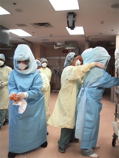
(2) Physical Methods
a) X-Ray Crystallography
In this method, X-rays are beamed at the crystal and electrons of the atoms of the sample being studied diffract the x-rays causing a diffraction pattern. Using mathematical formulae and computer programs 3-dimensional pictures and electron density maps can be created.
Since viruses are too small to be studied under a standard light-microscope, this method can be used to observe viruses.
b) Electron-Microscopy
Since viruses are too small to be observed in a light microscope, electron microscopy can be used to created detailed and accurate images of viruses. Electron-microscopy works by bombarding the specimen with a beam of electrons and the diffracted electrons can be used to form a image by using sensors connected to a computer that displays an image.
There are 2 types of electron microscopes:
· Transmission electron microscope (TEM)
· Scanning Electron microscope (STEM)
The image below shows the image produced by a TEM.
The image below shows the image produced by a STEM.
c) Ultra-centrifugation
Utra-centrifugation can be used to separate marcomolecules from each other when studying specimens such as cells or viruses. This enables researchers to obtain a purified sample of viruses.
Other function of ultra-centrifugation when using an analytical ultra-centrifuge are
A picture of a ultra-centrifuge is shown below.
(3) Serological methods/Immunological methods
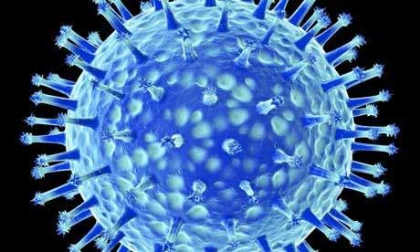
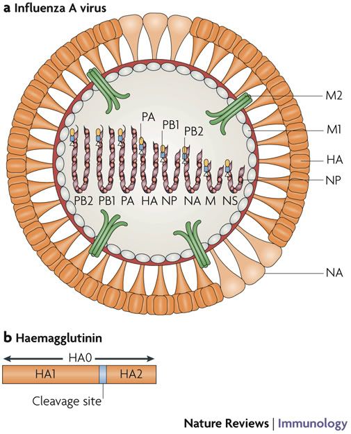
a) Haemagglutination (HA)
Certain types of viruses such as the influenza virus contain spikes on their envelope which are called:
· Haemaglutanin
· Neuraminidase
The haemagglutanin spike binds to specifically to red blood cells bringing them together form clumps of red blood cells which will float to the surface of the liquid. Hence the no. of viruses can be enumerated by observing the amount of red blood cells clumped at the surface.
b) Haemagglutination Inhibitation (HI)
A clinical lab test used to detect the presence of a certain haemagglutinating virus or other haemagglutinin antigen based on whether the red blood cells in the sample lose the ability to clump together when the antibody to the virus or other antigen is added to it.
Haemagglutination Inhibition uses the same principle as ordinary Haemagglutination except that it uses antibody inhibition which binds to the virus neutralising it and hence preventing it from binding to red blood cells causing agglutination.
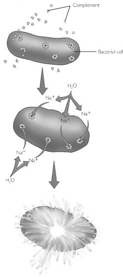
c) Complement Fixation
Complement fixation a a method where by an antibody binds to an antigen causing a complement cascade of molecules in the blood serum which interacts with the cell or pathogen causing it to lyse.
d) Immunostaining
Immunofluorescence
An antibody is tagged with fluorescent dye which attaches to a specific antigen of a sample. The sample is then observed under a fluorecent microscope which will produce exciting light to illuminate the fluorescent dye. This method can be used to study where and how the antibody and antigens react and bind together.
Immunogold Electron microscopy
Utilising the same principle in immunofluorescent staining except that flourescent dye is replaced with gold, antibody and antigen complexes can be observed in electron microscope where the gold dust can be seen.
e) Immunoprecipitation / Immunoblot
Immunoprecipitation
In this method a radioactively labelled antigen is reacted with an antibody creating a complex. This complex is will be run through SDS-PAGE and detected using an X-ray film. The diagram below furher explains this concept.
Immunoblot
A laboratory procedure, such as Western blot analysis, in which proteins that have been separated by electrophoresis are transferred onto nitrocellulose sheets and are identified by their reaction with labeled antibodies.
In this western blot analysis, the whole protein to be studied is run through SDS-PAGE and blotted onto nitrocellulose paper. Antibodies labelled with an indicator added to the paper, bind to the proteins on it. The paper is then treated with streptavidin which reacts with the indicator producing a colour reaction.

This diagram shows how the western blot is done. But in the case above radioactively labelled antibodies are used.
f) ELISA
ELISA also known as enzyme-linked immunosorbent assay is a biocemical technique used in immunology in the detection or antigens or antibodies that may be present in the sample.
One way the ELISA can work is by allowing an antibody to bind to the antigen on the well’s surface if the correct antibody-antigen pair is present. After which an enzyme conjugate added binds to the antibody-antigen complex, activatin the enzyme which converts an added substrate into a coloured compound. Any component missing will result in a negative test.
The diagram below explains this concept.
(4) Others and Molecular Biology
a) PAGE / SDS-PAGE
PAGE or Polyacrylamide Gel Electrophoresis is a method whereby proteins are separated by using an electric field to separate them according to their sizes in a polyacrylamide gel medium. The electric field produced pull the proteins through the ployacrylamide gel which is composed of a laberynth of tunnels within a mesh of fibres.
The SDS in SDS-PAGE is the step done before PAGE whereby the proteins are denatured so that it is reduced to its primary structure and that all proteins’ 3D shapes do not affect their rate of movement in the polyacrylamide gel in PAGE.
The SDS is actually an acronym for sodium dodecyl sulfate which is the detergent used to denature the proteins. The detergent dissolves hydrophoic molecules and has a negative charge attached to it. So if it is used on a protein, it dissolves it, denatures it and gives it negative charges. Hence in PAGE, proteins will migrate to the positive pole.
The diagram below shows what happens when proteins are degraded by SDS.
b) Western blot
This was discussed in the earlier section on immunoblot.
c) Protein Sequencing
In protein sequencing, the amino acid sequence on the poly peptide’s chain is determined by various methods.
To determine the composition of the protein the following can be done.
Another method in protein sequence is mass spectrometry where by the specific amino acid sequences can be determined. Mass spectrometry work by ionising the sample to created charged ions where their mass to charge ratio is determined. This ratio is calculated from the motion of the ions as they pass through electromagnetic fields.
The pictures below show a mass spectrometer and the data produced by it.
d) X-ray Crystallography
This was discussed earlier under the physical methods section.
e) Restriction Analysis
In the restriction analysis method the genetic material for example DNA is cut into segments using restriction enzymes and run through PAGE or gel electrophoresis separating the segments which can be analysed.
f) DNA Sequencing
In DNA sequencing, a mixture of bases, enzymes and cofactors are reacted with a DNA strand to create a complementary strand which can be run through gel electrophoresis. Through gel electroporesis the DNA bases can be sorted according to sequence and base pair type. By comparing the results with complementary bases, the sequence of the sample DNA strand can be recontructed.
g) Southern blot
Southern blot is a technique developed for transferring DNA fragments from gel electrophoresis onto the nitrocellulose paper. This is essential so that the DNA fragments can be detected using techniques such as labelling fragments with radioactive material.
The picture below shows how sourthern blotting is done.
h) Northen Blot
The northern blot analysis is based on the same principle as with the southern blot except that northern blot is used for RNA instead.
i) PCR / RT-PCR
PCR
PCR also known as Polymerase Chain Reaction is used to create a large amount of copies of DNA. This is useful especially when the DNA sample being studied is low in quantity.
PCR works by repeating a cycle of 3 steps over and over again till enough duplicate DNA is produced. The steps are:
RT-PCR
RT-PCR is used for RNA and works almost the same as standard PCR only with a an additonal step.
RT-PCR works by first converting RNA into DNA using reverse transcriptase to produce a complementary strand. Next the standard 3 step PCR cycle is used to create duplicates.
The video below shows how PCR works.
http://www.youtube.com/watch?v=j9Gu7iwBi4I
References:
Information:
1) http://pathmicro.med.sc.edu/mhunt/replicat.htm
2) http://www.stolaf.edu/people/hansonr/mo/x-ray.html
3) http://www.bio.davidson.edu/COURSES/GENOMICS/method/SDSPAGE/SDSPAGE.html


Attachment of HIV to a CD4+ cell. The outer domain of gp120 binds to the CD4 antigen. This leads to a conformational change in gp120 and a co-receptor binding site is exposed. This region of gp120 binds to the chemokine receptor. Binding to the chemokine receptor allows another conformational change to occur so that regions of the gp41 HIV protein interact to form a fusion domain that allows the viral and cell membrane to fuse.
HIV life cycle
AIDS
AIDS- transmission
· Sexual contact
· Blood and blood products
· Mother to child
- Placenta
- Mucosa
- Breast milk
· Pandemic
Symptomatic Stage
There are 4 stages:
Stage 1: Primary HIV Infection
The first stage of HIV infection is called primary infection. Primary infection begins shortly after an individual first becomes infected with HIV. This stage lasts for a few weeks. During this time period, individuals experience symptoms similar to the flu. Very few individuals seek treatment during this time, and those who do are usually misdiagnosed with a viral infection.
Often, if an HIV test is performed, it will come back negative, since antibodies are not yet being produced by the individual’s immune system. Those who believe they have been exposed to HIV should repeat the test again after six months.
Stage 2: Asymptomatic HIV
In the second stage, individuals are free from any symptoms of HIV. Levels of HIV in the blood are very low, but are detectable. If an HIV test is performed, it will come back positive. While the individual is asymptomatic, the HIV in their blood is reproducing constantly. This stage lasts about ten years, but can be much longer or shorter depending on the individual.
Stage 3: Symptomatic HIV
In the third stage, the immune system has become so damaged by HIV that symptoms begin to appear. Symptoms are typically mild at first, and then slowly become more severe. Opportunistic infections, infections that take advantage of the immune system’s vulnerable state, begin to occur. These infections affect almost all the systems of the body and include both infections and cancers. Some common opportunistic infections include tuberculosis, cytomegalovirus, and shingles.
Stage 4: Acquired Immune Deficiency Syndrome
In the fourth and final stage, a person is diagnosed as having AIDS. To be diagnosed as having AIDS, a person has to exhibit certain opportunistic infections, such as HIV wasting syndrome, pneumocystis pneumonia, or Kaposi sarcoma. Once a person is diagnosed with AIDS, they can never return to a stage of HIV, even if the individual gets better.
AIDS-related complex
- Diseases not considered definitive of AIDS
- May be attributed to HIV infection
- Indicative of detect in cell-mediate
- Immunity
AIDS
- Opportunistic infections as a result of fall in CD4 lymphocytes
AIDS therapy
Non-specific therapeutic management
- To boost general health
- Vitamins
- Minerals
- Anti-oxidants
- Others
Specific therapeutic management:
- Antiretroviral therapy
Nucleoside Reverse transcriptase Inhibitors:
-AZT (azidothymidine)
-3TC (lamivudine)
Non-nucleoside Reverse Transcriptase Inhibitors
-Efavirenz
-Nevirapine
*Rapid mutations due to inefficiency of reverse transcriptase
Vaccines
-Many candidates under development and trails.
-None so far proven useful
Video:
How HIV become Aids.


![]()


West Nile Virus
· Originated in Uganda
· Discovered in 1937
· Common in Africa, west Asia, Europe, Middle East
· Mainly mild to no symptoms
· Fever, headache, body rashes, skin rash , swollen lymph glands
· Severe symptoms
§ Crossing blood brain barrier
§ Encephalitis
§ Meningitis
· Mainly in persons above 50 years
West Nile Virus Transmission Cycle
![]()


