


Plaque Assay
Plaque assay is a method based on the principle that one virus will infect one cell which will produce a visible plaque on a cell monolayer in a plaque assay.
In a plaque assay the following takes place:

The plaque assay despite being a very simple method is very time-consuming and can only work for cells that can infect monolayers and cause cell lysis.
(2) Physical Methods
a) X-Ray Crystallography
In this method, X-rays are beamed at the crystal and electrons of the atoms of the sample being studied diffract the x-rays causing a diffraction pattern. Using mathematical formulae and computer programs 3-dimensional pictures and electron density maps can be created.
Since viruses are too small to be studied under a standard light-microscope, this method can be used to observe viruses.


b) Electron-Microscopy
Since viruses are too small to be observed in a light microscope, electron microscopy can be used to created detailed and accurate images of viruses. Electron-microscopy works by bombarding the specimen with a beam of electrons and the diffracted electrons can be used to form a image by using sensors connected to a computer that displays an image.
There are 2 types of electron microscopes:
· Transmission electron microscope (TEM)
· Scanning Electron microscope (STEM)
The image below shows the image produced by a TEM.

The image below shows the image produced by a STEM.

c) Ultra-centrifugation
Utra-centrifugation can be used to separate marcomolecules from each other when studying specimens such as cells or viruses. This enables researchers to obtain a purified sample of viruses.
Other function of ultra-centrifugation when using an analytical ultra-centrifuge are
A picture of a ultra-centrifuge is shown below.

(3) Serological methods/Immunological methods
a) Haemagglutination (HA)
Certain types of viruses such as the influenza virus contain spikes on their envelope which are called:
· Haemaglutanin
· Neuraminidase
The haemagglutanin spike binds to specifically to red blood cells bringing them together form clumps of red blood cells which will float to the surface of the liquid. Hence the no. of viruses can be enumerated by observing the amount of red blood cells clumped at the surface.

b) Haemagglutination Inhibitation (HI)
A clinical lab test used to detect the presence of a certain haemagglutinating virus or other haemagglutinin antigen based on whether the red blood cells in the sample lose the ability to clump together when the antibody to the virus or other antigen is added to it.
Haemagglutination Inhibition uses the same principle as ordinary Haemagglutination except that it uses antibody inhibition which binds to the virus neutralising it and hence preventing it from binding to red blood cells causing agglutination.
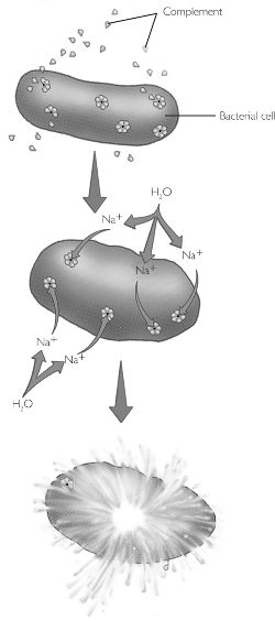
c) Complement Fixation
Complement fixation a a method where by an antibody binds to an antigen causing a complement cascade of molecules in the blood serum which interacts with the cell or pathogen causing it to lyse.
d) Immunostaining
Immunofluorescence
An antibody is tagged with fluorescent dye which attaches to a specific antigen of a sample. The sample is then observed under a fluorecent microscope which will produce exciting light to illuminate the fluorescent dye. This method can be used to study where and how the antibody and antigens react and bind together.

Immunogold Electron microscopy
Utilising the same principle in immunofluorescent staining except that flourescent dye is replaced with gold, antibody and antigen complexes can be observed in electron microscope where the gold dust can be seen.
e) Immunoprecipitation / Immunoblot
Immunoprecipitation
In this method a radioactively labelled antigen is reacted with an antibody creating a complex. This complex is will be run through SDS-PAGE and detected using an X-ray film. The diagram below furher explains this concept.

Immunoblot
A laboratory procedure, such as Western blot analysis, in which proteins that have been separated by electrophoresis are transferred onto nitrocellulose sheets and are identified by their reaction with labeled antibodies.
In this western blot analysis, the whole protein to be studied is run through SDS-PAGE and blotted onto nitrocellulose paper. Antibodies labelled with an indicator added to the paper, bind to the proteins on it. The paper is then treated with streptavidin which reacts with the indicator producing a colour reaction.

This diagram shows how the western blot is done. But in the case above radioactively labelled antibodies are used.
f) ELISA
ELISA also known as enzyme-linked immunosorbent assay is a biocemical technique used in immunology in the detection or antigens or antibodies that may be present in the sample.
One way the ELISA can work is by allowing an antibody to bind to the antigen on the well’s surface if the correct antibody-antigen pair is present. After which an enzyme conjugate added binds to the antibody-antigen complex, activatin the enzyme which converts an added substrate into a coloured compound. Any component missing will result in a negative test.
The diagram below explains this concept.

(4) Others and Molecular Biology
a) PAGE / SDS-PAGE
PAGE or Polyacrylamide Gel Electrophoresis is a method whereby proteins are separated by using an electric field to separate them according to their sizes in a polyacrylamide gel medium. The electric field produced pull the proteins through the ployacrylamide gel which is composed of a laberynth of tunnels within a mesh of fibres.
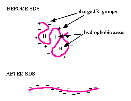
The SDS in SDS-PAGE is the step done before PAGE whereby the proteins are denatured so that it is reduced to its primary structure and that all proteins’ 3D shapes do not affect their rate of movement in the polyacrylamide gel in PAGE.
The SDS is actually an acronym for sodium dodecyl sulfate which is the detergent used to denature the proteins. The detergent dissolves hydrophoic molecules and has a negative charge attached to it. So if it is used on a protein, it dissolves it, denatures it and gives it negative charges. Hence in PAGE, proteins will migrate to the positive pole.
The diagram below shows what happens when proteins are degraded by SDS.
b) Western blot
This was discussed in the earlier section on immunoblot.
c) Protein Sequencing
In protein sequencing, the amino acid sequence on the poly peptide’s chain is determined by various methods.
To determine the composition of the protein the following can be done.
Another method in protein sequence is mass spectrometry where by the specific amino acid sequences can be determined. Mass spectrometry work by ionising the sample to created charged ions where their mass to charge ratio is determined. This ratio is calculated from the motion of the ions as they pass through electromagnetic fields.
The pictures below show a mass spectrometer and the data produced by it.


d) X-ray Crystallography
This was discussed earlier under the physical methods section.
e) Restriction Analysis
In the restriction analysis method the genetic material for example DNA is cut into segments using restriction enzymes and run through PAGE or gel electrophoresis separating the segments which can be analysed.
f) DNA Sequencing
In DNA sequencing, a mixture of bases, enzymes and cofactors are reacted with a DNA strand to create a complementary strand which can be run through gel electrophoresis. Through gel electroporesis the DNA bases can be sorted according to sequence and base pair type. By comparing the results with complementary bases, the sequence of the sample DNA strand can be recontructed.
g) Southern blot
Southern blot is a technique developed for transferring DNA fragments from gel electrophoresis onto the nitrocellulose paper. This is essential so that the DNA fragments can be detected using techniques such as labelling fragments with radioactive material.
The picture below shows how sourthern blotting is done.

h) Northen Blot
The northern blot analysis is based on the same principle as with the southern blot except that northern blot is used for RNA instead.
i) PCR / RT-PCR
PCR
PCR also known as Polymerase Chain Reaction is used to create a large amount of copies of DNA. This is useful especially when the DNA sample being studied is low in quantity.
PCR works by repeating a cycle of 3 steps over and over again till enough duplicate DNA is produced. The steps are:
RT-PCR
RT-PCR is used for RNA and works almost the same as standard PCR only with a an additonal step.
RT-PCR works by first converting RNA into DNA using reverse transcriptase to produce a complementary strand. Next the standard 3 step PCR cycle is used to create duplicates.
The video below shows how PCR works.
http://www.youtube.com/watch?v=j9Gu7iwBi4I
References:
Information:
1) http://pathmicro.med.sc.edu/mhunt/replicat.htm
2) http://www.stolaf.edu/people/hansonr/mo/x-ray.html
3) http://www.bio.davidson.edu/COURSES/GENOMICS/method/SDSPAGE/SDSPAGE.html